The hybrid operating theatres at Clinique Cecil, Klinik Im Park and Klinik Hirslanden enable precise imaging diagnostics during surgery.
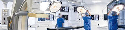
Clinique Cecil, Klinik Im Park and Klinik Hirslanden each have an ultra-modern hybrid operating theatre. The high-tech equipment enables surgeons to make diagnoses during operations based on precise imaging and to perform either open or minimally invasive surgery or a combination of both. For the patient, this means a faster, safer and less burdensome operation.
The hybrid operating theatre greatly increases the range of possible vascular procedures. Particularly older and seriously ill patients with complex vascular diseases benefit from the high-tech combination of a comprehensively equipped operating theatre with sterile clean room conditions and a high-performance imaging system that can be positioned around the patient in virtually all directions and angles. The system can generate high-precision 3D images of the vessels and enables computer-aided navigation and orientation during the operation.
New techniques, greater teamwork
The key advantage of the hybrid operating theatre is that it can be used to perform open and minimally invasive surgery, or a combination of both, while providing high-precision radiological imaging during the procedure. This paves the way for entirely new interdisciplinary therapeutic concepts that are faster, safer and less traumatic for patients, because everything can be carried out simultaneously in one place.
Particularly in the case of vascular emergencies, it is possible to convert a minimally invasive operation into an open operation in just a few minutes, without the patient having to be moved to another theatre at such a critical time. Various examinations can also be incorporated into the procedure. For instance, the surgical team can measure and evaluate the patient’s blood pressure, blood flow rate, the amount of blood being transported through the heart and the elasticity of the vessels. This saves the patient from undergoing time-consuming individual examinations, or being transported to various diagnostic devices during the operation.
Setting up a hybrid operating theatre is a very challenging task: not only does the imaging unit require additional space, but such theatres are also staffed by more personnel. Depending on the complexity and difficulty of the operation, a team of up to 20 people are involved, including anaesthetists, vascular surgeons, endovascular specialists, operating theatre nurses , cardiology technicians and supporting personnel such as experts from implant manufacturers.
High-precision imaging during the operation
The centrepiece of the hybrid operating theatre is the modern angiography unit, offering several advantages. Guided by a computer, it rotates around the patient on a robotic arm with eight degrees of freedom and generates images from every possible angle. The three-dimensional images are immediately available in excellent quality on monitors in the operating theatre.
Surgeons can therefore use the angiography system to navigate their way inside the body, making it easier for them to perform complex procedures. Unlike conventional mobile x-ray devices with angiographic capabilities, which are manually moved into a specific position next to the patient (known as C-arms), surgeons can use a computer to precisely position both the operating table and a robotic arm featuring the curved x-ray apparatus. If necessary, the modern flat panel x-ray detector can rotate around the patient so quickly that the image quality and definition is comparable to that of computer tomography (CT). The device also requires a lower dose of radiation than the previous models, which is beneficial for the patient and the operating theatre team.
The computer uses the image data to generate a three-dimensional representation of the application area in close to real time. Based on this virtual model, surgeons use a specialised computer programme to verify the best approach just before the actual operation begins and also receive a kind of route plan for the catheter or surgery. During the procedure, the imaging makes it possible to instantaneously review the progress of the operation.
The operating table is directly connected to the angiography unit, so the computer can record the exact position of the table in the room at all times, which is a significant advantage for the surgical navigation. The angle and height of the table can also be adjusted to accommodate all the necessary patient positions, which is essential for achieving the best possible open surgery outcome, regardless of the organ or region of the body under operation.
Hybrid operating theatre in action: aortic aneurysms
These days aneurysms in the abdomen’s main artery (abdominal aortic aneurysms) are increasingly treated by inserting a vascular prosthesis inside the vessel, if this is possible given the patient’s anatomy (link only available in German and French). The prosthesis can prevent potentially life-threatening ruptures of the artery. During minimally invasive surgery, a vascular prosthesis with a metal lattice structure (stent) is inserted via the groin vessels into the main artery, where it is assembled and fixed in place using a blood-tight seal.
Thanks to the hybrid operating theatre’s angiography unit, the team can immediately check whether the stent is correctly positioned and the aneurysm has been entirely removed from the blood stream. This makes it possible to instantly verify the success of the surgery, which in turn shortens the duration of the operation. Vascular surgeons not only use the hybrid angiography system to ensure the precise positioning of the stent – they also use the same software to plan the operation.
Due to repositioning of the patient and the insertion of inflexible instruments the patient’s anatomy changes between the pre-operative CT scan and the radioscopy carried out during surgery. That is why the use of 3D surgical images makes the planning and implantation of prostheses even more precise. The images automatically subdivide the aorta and markers can be placed on its 3D form to highlight certain aspects such as the outflow of the kidney arteries. These markings are then overlaid onto the shape of the aorta during the live radioscopy. If any changes are made to the position of the operating table, the C-arm or the angle of the C-arm then the superimposed image is adjusted accordingly. The advantages of the hybrid operating theatre are especially significant when it comes to complex vascular prostheses.
Article from Prof.Dr.med. Hardy Schumacher, surgeon specialising in vascular surgery.