
Melanoma is the fifth most common cancer overall, and the most malignant form of skin cancer. The malignant tumour usually appears as a black, irregular spot on the skin and, unlike non-melanoma skin cancer, sometimes appears before the age of 50. Since it can be aggressive, metastasise and attack other tissues, early diagnosis and treatment is particularly important.
Overview of melanoma skin cancer
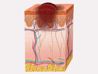
Melanoma skin cancer, also known as (malignant) melanoma, occurs in the basal cell layer of the epidermis. It develops slowly – over months or years – from melanocytes, the skin’s pigment-producing cells, which make the pigment melanin and are therefore responsible for tanning the skin.
If melanocytes degenerate, these pigment cells begin to proliferate uncontrollably. This is melanoma. As soon as melanoma develops, it becomes visible on the epidermis. Less commonly, it arises from an existing mole or dark birthmark, or in the eyes, mucous membranes or brain.
Melanoma skin cancer is generally divided into different stages, which indicate the progression of the disease. The different stages provide information on how far a tumour has already spread, whether lymph nodes have already been affected and whether metastases have formed. The sooner melanoma is detected and removed, the better the chances of recovery.
If patients wait, cancer cells may enter lymph nodes or internal organs via the lymphatic system or bloodstream, where they eventually form further tumours (metastasise). The risk of metastases depends on the thickness of the melanoma. The deeper it penetrates, the sooner metastases may occur.
Melanoma skin cancer: melanoma types
Malignant melanoma has many faces: its diagnosis and treatment depend on the symptoms. The early detection and correct classification of melanomas is essential for successful treatment. Early detection of skin cancer through appropriate examination of moles can significantly improve the prognosis for patients.
Melanomas are divided into the following types:
- Superficial spreading melanoma (SSM): As it initially spreads only over the surface of the skin, there is a good chance of recovery if it is diagnosed early.
- Nodular melanoma (NM): Nodular melanoma is characterised by dark, raised nodes on the skin surface. This is an aggressive type of tumour, which develops rapidly, penetrates the skin early on, and leads to the formation of metastases. It is usually only discovered at an advanced stage.
- Lentigo-maligna melanoma (LMM): This melanoma, which mainly occurs in older people, develops slowly from lentigo maligna skin patches that have been exposed to the sun over a long period of time. This typically flat melanoma often, but not exclusively, occurs on the face, and is usually dark in colour.
- Acral lentiginous melanoma (ALM): This rare form of melanoma appears as a dark streak or patch and is often found on the palms of the hands, the soles of the feet and under nails.
- Amelanotic melanoma: Amelanotic melanoma is a rare but aggressive form of melanoma skin cancer that is particularly challenging to identify. It shows little or no pigmentation, often appears pink, reddish or colourless and therefore often gives the appearance of other skin conditions.


Melanoma skin cancer: causes and risk factors
Like non-melanoma skin cancers, malignant melanoma forms as a consequence of overly strong or intense UV radiation. Prolonged or unprotected periods in the sun damage the skin, as does strong radiation from artificial sources such as sunbeds. UV radiation can damage and alter the genetic material in melanocytes, which can lead to unchecked cell proliferation and the development of melanoma.
Some people have a particularly high risk of developing melanoma skin cancer. People with pale skin are more at risk of developing malignant melanoma. People with many moles (more than 100 moles in total) are also at greater risk. In particular, many moles showing a mix of colours (dysplastic moles) or of conspicuously large size indicate an increased risk.
A family history of melanoma also increases the risk. In addition, your own immune system and how well you were protected against the sun during childhood and adolescence also play an important role in assessing the risk of skin cancer. Anyone who suffered severe sunburns in childhood and adolescence, has already suffered from melanoma or has a weakened immune system, for example after an organ transplant or in the case of an immune-impairing illness such as HIV, must also expect a higher risk of melanoma skin cancer.
Symptoms of melanoma skin cancer
Melanoma usually appears as an irregular black mark on the skin. These can vary considerably in appearance, from small and flat to large and raised. However, in contrast to non-melanoma skin cancers, melanoma can form not only in sun-exposed areas, but on any part of the body. The back, legs or face are often affected. In rare cases, however, the cancer can also occur on fingernails, mucous membranes or other parts of the body, including the scalp.
In addition to the dark colouration of the skin, malignant melanoma can also itch and bleed in later stages, and may be associated with poor wound-healing. Pain is unusual. If a mole turns malignant, the first warning signs are spontaneous bleeding, a red border forming around the edges, or changes in shape or colour. Symptoms like these should be taken seriously, and should be examined promptly by a dermatologist.
Patients may also suffer from various concomitant symptoms, which usually occur at an advanced stage of the illness. These include fever, night sweats and unintended weight loss.
Diagnosing melanoma skin cancer
To determine the type of pigmented skin lesion, the specialist examines the spot with the aid of a dermatoscope or epiluminescence microscope. This enables them to more accurately identify the structure and distinguish it from a normal mole. Finally, the ABCDE rule can be used to diagnose it:
- A: Asymmetry → The patch is asymmetrical, not circular
- B: Border irregularity → The mark is not sharply demarcated from its surroundings
- C: Colour variation → The skin patch is a mix of different colours such as brown, black, pink or even purple, and/or colour changes
- D: Diameter → The diameter is larger than 5 millimetres
A further parameter may be used to give additional information following long-term observation: - E: Evolving → The skin spot gets bigger over time
The ABCDE rule can also help you with self-examination. As a general rule, moles that change in terms of colour, shape or size should be examined by a dermatologist as soon as possible. This also applies to itchy moles, moles that bleed easily from minor injuries, or moles that are conspicuous in other ways. These should, without exception, be examined by a doctor.
If one or more of the ABCDE criteria are met, malignant melanoma is possible. After an initial assessment, the thickness of the tumour is measured with a special ultrasound device. The next step is to determine how deeply it has penetrated the skin and whether it has already metastasised.
The tissue sample taken during a biopsy is then examined in the laboratory. If malignant melanoma is diagnosed, the attending doctor will draw up an individual treatment plan.
Treatment of melanoma skin cancer
There are a variety of treatment options for melanoma skin cancer. The aim of treatment is always the complete removal of the cancer. To achieve this, sometimes several treatment measures are used in combination.
Surgical therapy
The first step is to surgically remove the melanoma along with a little of the surrounding tissue as a safety margin, to guard against a return of the disease. After the excision, the tumour is sent to the laboratory to be examined and checked for signs of radial growth. If these are present and extend to the edge of the area that has been cut out, then more tissue needs to be removed from that area.
For smaller and easily accessible tumours, a dermatologist will perform the surgery. Larger interventions are carried out by specialist surgeons. Plastic surgery will also be involved if any skin grafts are required.
After removal of the tumour, it is measured and analysed in the laboratory. If it is thicker than one millimetre and without any visible signs of spreading, then a biopsy of the ‘sentinel’ lymph nodes (those nearest to the tumour) can indicate whether cancer cells have spread from the melanoma into other organs. The reasoning is that the cancer cells would have had to pass through these lymph nodes and would therefore be detectable in its tissue. The result is key to deciding on the course of further treatment.
Adjuvant therapy
Whenever possible, melanoma skin cancer is treated surgically. Depending on its size, position, spread and the health of the patient, subsequent adjuvant (supportive) therapy may also be useful. It is used when the tumour has been completely removed but when there is still a risk of recurring melanoma. The aim of this therapy is to achieve a long-term cure by preventing new tumours from forming and by destroying any undetected metastases.
Adjuvant measures include:
- medication therapy
- radiotherapy
Medication therapy
If the tumour has spread to other organs, there are various groups of medication that can destroy the cancer cells in the body.
- Immunotherapy: Immunotherapy involves prompting the body to attack the cancer cells itself. The diseased cells are either marked so that the immune system can recognise them and take action against them, or the medication actively triggers a defence response against the cancer cells.
- Targeted drugs: Certain drugs work in the affected cells themselves. With targeted treatment, they can attack the signalling structures of the cancer cells and thus inhibit their growth.
- Chemotherapy: Chemotherapy uses what are known as cytostatic drugs to hinder the growth and division of cancer cells in the body. As a result, tumours become smaller or break up completely.
Radiotherapy
If the melanoma has formed metastases in the brain, lymph nodes, bones or other areas that are sensitive or hard to access, then the tumour can be treated with radiotherapy that is gentle on the surrounding tissue.
Your attending specialist will inform you about all the opportunities and risks of the various forms of treatment in an individual consultation.
Prevention
There are several risk factors that can lead to the development of skin cancer, and to its growth and spread. Prevention of malignant melanoma requires careful and consistent sun protection. The following measures serve to prevent melanoma skin cancer:
- Always apply a sun cream that protects you from both UVA and UVB radiation, even on cloudy days.
- If you will be spending long periods outdoors, wear long clothing that covers the skin, especially sensitive areas.
- If possible, avoid being outdoors in the strongest sunlight (between 11 am and 3 pm).
- Avoid sunbathing and, wherever possible, try to stay in the shade.
The UV damage that leads to melanoma often originates in childhood or adolescence. This means that moles should always be kept under close observation and any changes in the skin should always be checked by a specialist doctor. If it is found early, melanoma can be successfully treated and spreading can be prevented.
Should patients who themselves suspect that they might have skin cancer undergo a medical examination?
If a patient finds superficially visible pigmented marks on the skin that look unusual or develop noticeably, for example in the form of a change in colour or rapid growth, these should be checked by a specialist.
Is melanoma skin cancer dangerous?
Yes, it is a malignant illness that can be very aggressive if left untreated. The tumour can penetrate deeper layers of the skin and lead to metastases. Early detection and rapid treatment are the be-all and end-all for a good prognosis.
Can melanoma skin cancer be cured if it has spread?
If cancer has already formed metastases, the illness is more difficult to treat, but medical innovations such as immunotherapy can still fight it in a targeted manner. The treatment is individually tailored and can have a positive effect on the course of the disease.